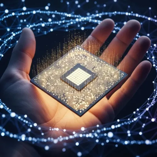
Physics in cancer diagnostics is changing lives. As we have often heard, Precision, and accuracy are crucial in the treatment of cancer. Especially with PET-MRI imaging, it is possible to find a tumor’s precise situation. Doctors can plan radiation in a way that treats cancer and spares as much healthy tissue as possible. Modern cancer treatments make full use of physics, which is a basic area of scientific research. Moreover, modern technologies use physics to find tumors. They look at their behavior and come up with new treatments. Physics applications allow imaging inside the body. They target cancerous cells. These breakthroughs in cancer diagnostics and treatment are revolutionizing radiation oncology.
Core physics principles are contributing a lot to breakthroughs in all aspects of cancer diagnosis. Such efforts also use innovative imaging techniques like positron emission tomography (PET) and magnetic resonance imaging (MRI) to direct treatment. In this article, we will cover radiation oncology, where radiation is delivered powerfully and precisely for radiation safety.
Physics in Cancer Diagnostics: Making Scans Work
Just like finding hidden things in a maze can be challenging, similarly detecting tumors inside the body is equally difficult. Doctors use advanced technologies to detect these cancers without needing to open the body. Physics helps make these tools. It’s the science of how things work in the world, like light, magnets, and tiny particles.
Physics helps the doctors to diagnose where cancer might be. It helps them to understand how different parts of the body look using special pictures. Two big ways physics helps are with PET scans and MRI scans. These are like super-powered cameras that show what’s happening inside our body.

Look at this picture. It shows how PET and MRI use different ideas from physics to make treatments. They help doctors make good plans to treat cancer.
1. Positron Emission Tomography (PET)
Researchers use Positron Emission Tomography as a functional tool in imaging to offer a detailed analysis of activities in the metabolism of tissues. The physics starts with the radiotracer; a molecule, which bears radioisotope labeled for positron-emitting radionuclide. When a radiotracer is introduced into a body, it will accumulate in those regions which are metabolically active. For example, fast-growing cancer cells. The emitted positron travels a short distance. It then annihilates with an electron. This produces two gamma rays that are ejected and travel in opposite directions.
PET scanners detect these coincident gamma rays and complex algorithms, drawing from physics principles of radiation detection and reconstruction, reconstruct the origin of the annihilation event. This enables clinicians to quantify metabolic uptake in various regions. The nuclear medicine physics informs the design of these radiotracers and the conceptualization of their half-lives. See examples of common radiotracers and their properties in the table below.
| Radiotracer (Isotope) | Common Use Example (Molecule) | Half-life |
|---|---|---|
| Fluorine-18 (¹⁸F) | FDG (Glucose analog) | Approx. 110 mins |
| Carbon-11 (¹¹C) | Choline, Methionine | Approx. 20 mins |
| Nitrogen-13 (¹³N) | Ammonia | Approx. 10 mins |
| Oxygen-15 (¹⁵O) | Water | Approx. 2 mins |
| Gallium-68 (⁶⁸Ga) | DOTA compounds | Approx. 68 mins |
| Copper-64 (⁶⁴Cu) | Various targeting molecules | Approx. 12.7 mins |
2. Magnetic resonance imaging (MRI) Scans
Magnetic resonance imaging is a procedure to make pictures of the inside of our body. It does not use X-rays; although, it relies on magnets and radio waves. Our body is mostly made of water and water has small parts like magnets. The big magnet on the MRI machine makes these small parts line up in a single direction. Then, the machine transmits radio waves that send these small parts. When they return to their original position, they emit their own radio signals.
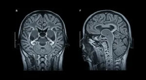
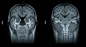
The MRI machine hears such signals. One body part will report back different from another body part. This makes the machine draw a clear picture. Doctors can view tumors in position and watch how water and blood flow from it, thus knowing the tumor better. The pictures are changing with a slider for you to see. When the radio waves are altered, the picture looks changed, and it allows the doctors to see more about the tumor. Such imaging techniques are of great help in treatment plans. It is all diagnostic radiology.
Latest Developments in Radiation Oncology
Doctors have to treat the cancer after finding it. Once doctors detect cancer, they have to cure it. Radiation oncology is a technique of treating cancer through a high form of energy like X-rays, or other small particles. The idea is to damage or destroy cancer cells; while leaving normal cells unharmed. Physics is very important to ensure that the energy reaches the right place, with the right force.
Physics assists doctors to point the energy as a superhero would to point a beam of laser. They use it to decide that the radiation dose is appropriate for the tumor. The infographic demonstrates how the energy enters the body.

Radiation treatment can be given using physics in two main forms.
1. External Beam Radiation Therapy (EBRT)
There is a primary category of radiation therapy known as external beam radiation therapy. An outside large machine beams energy to the tumor. The standard machine known is called a linear accelerator, or LINAC. These energy beams are manufactured using the same deep understanding of physics in cancer diagnostics that helps us see tumors so clearly.
Physics also designs the beams. It’s as if you shone a flashlight through a cut-out shape. Physics gives a shape to the energy beam such that only the tumor suffers the beam. This spares the healthy areas of the body and lowers side effects. Radiation protection is the prevention of the energy from harming others to keep everyone safe.
2. Nuclear Medicine Approaches
Nuclear medicine uses radioactive materials to help doctors see inside the body and, from time to time, also to treat some health problems. Y-90 or I-131 is bonded to molecules in targeted therapy. They connect to cancer cells and concentrate the radiation in the tumor. I-131 therapy is applied when treating patients after surgery for thyroid cancer. It helps clean up any bits of tissue that are left behind. There are strict rules in place to help ensure that people are protected from radiation. In neuroendocrine tumors, Lu-177 is used to search out certain receptors in the tumor and then kill the tumor cells using beta radiation. Physicists check the amount of radiation given to people to see that the right amount is used for treatment and not too much or too little.
| Radionuclide | Emission Type | Common Use |
|---|---|---|
| I-131 | Beta, Gamma | Thyroid cancer |
| Y-90 | Beta | Liver cancer |
| Lu-177 | Beta | Neuroendocrine tumors |
Integration into Treatment Plans
Clearing images from medical scans is important for planning the best treatments. From the pictures taken during PET or MRI scans, that are powered by physics in cancer diagnostics. Doctors see the size and exact location of the tumor. High-tech computer tools are used to process this data. Medical physicists use these programs. Radiation oncologists use mathematics to design the best way to target the tumor. They find the most effective and safest way to direct the radiation. Experts direct the radiation at the tumor while ensuring that the healthy nearby tissues are exposed to only a low amount of radiation. Both doctors and physicists collaborate as a team. As a result, the best treatment plan is created for each patient. The picture illustrates how imaging leads to treatment.

Keeping Everyone Safe from Radiation
Radiation therapy uses intense energy known as ionizing radiation. Scientists and medical staff should take measures to guarantee everyone’s safety. Physics shows us the ways in which energy interacts with the human body. Side effects can appear when radiation hits normal cells. The outcome depends on the radiation level. It also depends on the body area, for example, the head and neck.
Physics specifies safe radiation doses for all parts of the body. They implement measures like ALARA (As Low As Reasonably Achievable). This means they keep exposure to radiation as small as possible. Engineers apply physics to construct walls in treatment rooms. They also use it for regularly checking the machines for safety and accuracy. To guarantee radiation oncology treatment is safe, medical physicists invest a lot of time and effort. Keep in mind that strict safety rules and dedication to recommended doses at all times.
Real-Life Success Story
A patient diagnosed with pancreatic cancer was assisted by PET-MRI at a prestigious hospital. The scan showed how the cancer responded to medicine. Doctors used this information to decide if surgery was possible. The patient lived longer because of this. The table below show the results.
| Measure | Before Treatment | After Treatment |
|---|---|---|
| Tumor Size | 3.5 cm | 2.1 cm |
| PET signal | High | Low |
| Cancer Return | — | 15% |
Conclusion
The use of physics in cancer diagnostics and treatments make cancer prevention much better. Doctors can spot small cancer cells on the scans first and later use radiations to treat them. It supplies both equipment and information that help doctors detect and treat cancer. Recent developments in physics bring new opportunities to support and treat more people with cancer. Do you find science interesting? Want to be notified about similar stories? You can contact us for emails at Learning Breeze to enjoy more interesting science stories.